ClariCT.ai improves brain CT scans denoising in a multi-scanner study
- martapinto
- May 27, 2024
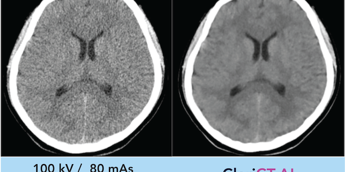
Significant improvements in diagnostic image quality for five CT scanners were achieve in this retrospective single-center German study by using a commercially available and FDA approved deep learning denoising (DLD) algorithm (ClariCT.AI, ClariPi, South Korea). Non-contrast cranial computed tomography images of 150 consecutive patients, who had undergone routine imaging after minor head trauma, were reconstructed using filtered back projection and a vendor-agnostic DLD method. DLD showed constantly superior results compared to FBP in overall diagnostic image quality, image noise, gray matter-white matter differentiation, artifacts, sharpness, and diagnostic confidence, as well as improvement in noise, signal-to-noise ratio, contrast-to-noise ratio and an artifact index for the posterior fossa. With the presented DLD algorithm, access to improved image quality would be possible for more than one scanner, scanners from different vendors, and older scanners in hospitals or radiological centers by making use of just one denoising engine.
Value of vendor-agnostic deep learning image denoising in brain computed tomography: A multi-scanner study
Rofo, 2024
Abstract
Objective: To evaluate the effect of a vendor-agnostic deep learning denoising (DLD) algorithm on diagnostic image quality of non-contrast cranial computed tomography (ncCT) across five CT scanners.
Materials and Methods: This retrospective single-center study included ncCT data of 150 consecutive patients (30 for each of the five scanners) who had undergone routine imaging after minor head trauma. The images were reconstructed using filtered back projection (FBP) and a vendor-agnostic DLD method. Using a 4-point Likert scale, three readers performed a subjective evaluation assessing the following quality criteria: overall diagnostic image quality, image noise, gray matter-white matter differentiation (GM-WM), artifacts, sharpness, and diagnostic confidence. Objective analysis included evaluation of noise, contrast-to-noise ratio (CNR), signal-to-noise ratio (SNR), and an artifact index for the posterior fossa.
Results: In subjective image quality assessment, DLD showed constantly superior results compared to FBP in all categories and for all scanners (p<0.05) across all readers. The objective image quality analysis showed significant improvement in noise, SNR, and CNR as well as for the artifact index using DLD for all scanners (p<0.001).
Conclusion: The vendor-agnostic deep learning denoising algorithm provided significantly superior results in the subjective as well as in the objective analysis of ncCT images of patients with minor head trauma concerning all parameters compared to the FBP reconstruction. This effect has been observed in all five included scanners.