Company: Brainreader Product: Neuroreader
Diagnostic utility of brain MRI volumetry in comparing traumatic brain injury, Alzheimer disease, and behavioral variant frontotemporal dementia
BMC Neurology, 2024
Abstract
Background
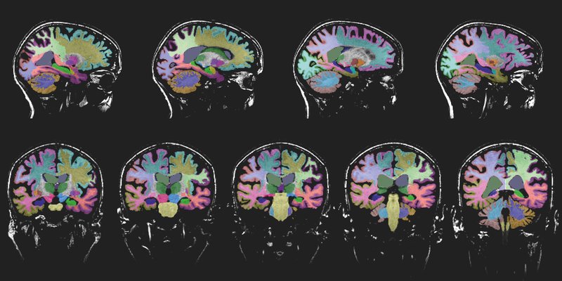
Brain MRI with volumetric quantification, MRI volumetry, can improve diagnostic delineation of patients with neurocognitive disorders by identifying brain atrophy that may not be evident on visual assessments.
Objective
To investigate diagnostic utility of MRI volumetry in traumatic brain injury (TBI), early-onset Alzheimer disease (EOAD), late-onset Alzheimer disease, and behavioral variant frontotemporal dementia (bvFTD).
Method
We utilized 137 participants of TBI (n = 40), EOAD (n = 45), LOAD (n = 32), and bvFTD (n = 20). Participants had 3D T1 brain MRI imaging amendable to MRI volumetry. Scan volumes were analyzed with Neuroreader. One-way ANOVA compared brain volumes across diagnostic groups. Discriminant analysis was done with leave-one-out cross validation on Neuroreader metrics to determine diagnostic delineation across groups.
Result
LOAD was the oldest compared to other groups (F = 27.5, p < .001). There were no statistically significant differences in sex (p = .58) with women comprising 54.7% of the entire cohort. EOAD and LOAD had the lowest Mini-Mental State Exam (MMSE) scores compared to TBI (p = .04 for EOAD and p = .01 for LOAD). LOAD had lowest hippocampal volumes (Left Hippocampus F = 13.1, Right Hippocampus F = 7.3, p < .001), low white matter volume in TBI (F = 5.9, p < .001), lower left parietal lobe volume in EOAD (F = 9.4, p < .001), and lower total gray matter volume in bvFTD (F = 32.8, p < .001) and caudate atrophy (F = 1737.5, p < .001). Areas under the curve ranged from 92.3 to 100%, sensitivity between 82.2 and 100%, specificity of 78.1-100%. TBI was the most accurately delineated diagnosis. Predictive features included caudate, frontal, parietal, temporal lobar and total white matter volumes.
Conclusion
We identified the diagnostic utility of regional volumetric differences across multiple neurocognitive disorders. Brain MRI volumetry is widely available and can be applied in distinguishing these disorders.
Read full study