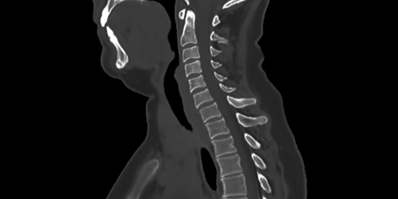
MRI-based synthetic CT offers a reliable radiation-free alternative for cervical spine fracture evaluation
Product: BoneMRI Company: MRIguidance
Radiological evaluation and clinical implications of deep learning- and MRI-based synthetic CT for the assessment of cervical spine injuries
European Radiology, 2025
Abstract
Objective
Efficient evaluation of soft tissues and bony structures following cervical spine trauma is critical. We sought to evaluate the diagnostic validity of magnetic resonance imaging (MRI)-based synthetic CT (sCT) compared with conventional computed tomography (CT) for cervical spine injuries.
Methods
In a prospective, multicenter study, patients with cervical spine injuries underwent CT and MRI within 48 h after injury. A panel of five clinicians independently reviewed the images for diagnostic accuracy, lesion characterization (AO Spine classification), and soft tissue trauma. Fracture visibility, anterior (AVH) and posterior wall height (PVH), vertebral body angle (VBA), segmental kyphosis (SK), with corresponding interobserver reliability (intraclass correlation coefficients (ICC)) and intermodal differences (Fleiss' Kappa), were recorded. The accuracy of estimating Hounsfield unit (HU) values and mean cortical surface distances were measured.
Results
Thirty-seven patients (44 cervical spine fractures) were enrolled. sCT demonstrated a sensitivity of 97.3% for visualizing fractures. Intermodal agreement regarding injury classification indicated almost perfect agreement (κ = 0.922; p < 0.001). Inter-reader ICCs were good to excellent (CT vs. sCT): AVH (0.88, 0.87); PVH (0.87, 0.88); VBA (0.78, 0.76); SK (0.77, 0.93). Intermodal agreement showed a mean absolute difference of 0.3 mm (AVH), 0.3 mm (PVH), 1.15° (VBA) and 0.51° (SK), respectively. MRI visualized additional soft tissue trauma in 56.8% of patients. Voxelwise comparisons of sCT showed good to excellent agreement with CT in terms of HUs (mean absolute error of 20 (SD ± 62)) and a mean absolute cortical surface distance of 0.45 mm (SD ± 0.13).
Conclusion
sCT is a promising, radiation-free imaging technique for diagnosing cervical spine injuries with similar accuracy to CT.
Key points
Question Assessing the accuracy of MRI-based synthetic CT (sCT) for fracture visualization and classification in comparison to the gold standard of CT for cervical spine injuries. Findings sCT demonstrated a 97.3% sensitivity in detecting fractures and exhibited near-perfect intermodal agreement in classifying injuries according to the AO Spine classification system. Clinical relevance sCT is a promising, radiation-free imaging modality that offers comparable accuracy to CT in visualizing and classifying cervical spine injuries. The combination of conventional MRI sequences for soft tissue evaluation with sCT reconstruction for bone visualization provides comprehensive diagnostic information.