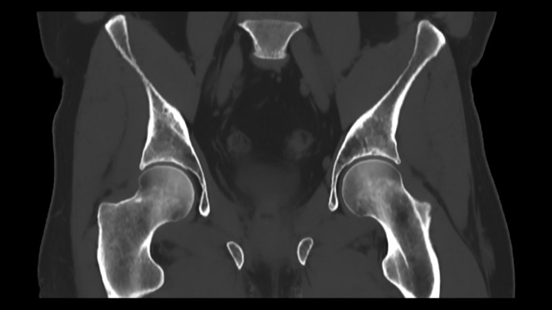
A U.S. prospective study investigated the effectiveness of BoneMRI (MRIguidance) in generating MRI-based synthetic CT (sCT) images compared to conventional CT (cCT) for evaluating pediatric hip disorders. The study included MRI scans from 38 hips of 19 patients with conditions like Developmental Dysplasia of the Hip and Femoroacetabular Impingement. The aim was to assess if sCT could serve as a viable alternative to cCT, potentially reducing radiation exposure in children. Results showed that measurements between sCT and cCT were statistically equivalent, with a mean surface distance between sCT and cCT at 0.23 mm ± 0.18 mm, demonstrating high agreement. Intraclass correlation coefficients (ICC) ranged from 0.79 to 0.99 for various hip morphometric parameters, indicating good to excellent reliability and agreement in measurements. These outcomes highlight sCT's promise for pediatric hip disorder evaluation, offering an alternative to traditional CT imaging.
Read full study
Abstract
Background
MRI-based synthetic CT (sCT) generates CT-like images from MRI data.
Objective
To evaluate equivalence, inter- and intraobserver reliability, and image quality of sCT compared to conventional (cCT) for assessing hip morphology and maturity in pediatric patients.
Materials and methods
We prospectively enrolled patients <21 years old with cCT and 3T MRI of the hips/pelvis. A dual-echo gradient-echo sequence was used to generate sCT via a commercially available post-processing software (BoneMRI v1.5 research version, MRIguidance BV, Utrecht, NL). Two pediatric musculoskeletal radiologists measured seven morphologic hip parameters. 3D surface distances between cCT and sCT were computed. Physeal status was established at seven locations with cCT as reference standard. Images were qualitatively scored on a 5-point Likert scale regarding diagnostic quality, signal-to-noise ratio, clarity of bony margin, corticomedullary differentiation, and presence and severity of artifacts. Quantitative evaluation of Hounsfield units (HU) was performed in bone, muscle, and fat tissue. Inter- and intraobserver reliability were measured by intraclass correlation coefficients. The cCT-to-sCT intermodal agreement was assessed via Bland-Altman analysis. The equivalence between modalities was tested using paired two one-sided tests. The quality parameter scores of each imaging modality were compared via Wilcoxon signed-rank test. For tissue-specific HU measurements, mean absolute error and mean percentage error values were calculated using the cCT as the reference standard.
Results
Thirty-eight hips in 19 patients were included (16.6 ± 3 years, range 9.9–20.9; male = 5). cCT- and sCT‐based morphologic measurements demonstrated good to excellent inter- and intraobserver correlation (0.77<ICC<0.98). Measurements were statistically equivalent (P < 0.05), with average intermodal differences <2°. Mean surface distance between cCT and sCT was 0.23 ± 0.18 mm. Accuracy to determine physeal maturity status by sCT was 98.4% (242/246, 95% CI 96.0–99.6). No significant differences were found on overall diagnostic quality, bony margin clarity, corticomedullary differentiation, and artifacts between sCT and cCT; signal-to-noise ratio was higher on sCT (P < 0.001). HU were within reference values on both sCT and cCT.
Conclusion
sCT is equivalent to cCT for the assessment of hip morphology, physeal status, and radiodensity assessment in pediatric patients.