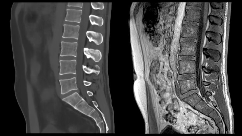
In this study, BoneMRI by MRIguidance was used as an alternative method to investigate a major unmet need for developing a sensitive assessment of structural damage in the spine in patients with axial spondyloarthritis (axSpA). The study compared the ability of BoneMRI generated MRI-based synthetic CT (sCT), low-dose CT (ldCT) and radiography to detect spinal new bone formation (NBF) in the spine of 17 patients with axSpA. The ldCT images were used as the standard reference. Results showed that sensitivity to detect NBF was much higher in MRI-based sCT (0.67) when compared to radiography (0.13), both with equally very high specificities (>0.95), when ldCT was considered the standard reference, despite limited training of the four scoring readers who participated in the study. This new method has the potential to markedly improve structural damage assessment in the spine of patients with axSpA, without any radiation exposure.
Read full story
MRI-based synthetic CT: a new method for structural damage assessment in the spine in patients with axial spondyloarthritis – a comparison with low-dose CT and radiography
Annals of the Rheumatic Diseases, 2024
Abstract
Objective: To investigate the ability of MRI-based synthetic CT (sCT), low-dose CT (ldCT) and radiography to detect spinal new bone formation (NBF) in patients with axial spondyloarthritis (axSpA).
Methods: Radiography of lumbar and cervical spine, ldCT and sCT of the entire spine were performed in 17 patients with axSpA. sCT was reconstructed using the BoneMRI application (V.1.6, MRIGuidance BV, Utrecht, NL), a quantitative three-dimensional MRI-technique based on a dual-echo gradient sequence and a machine learning processing pipeline that can generate CT-like MR images. Images were anonymised and scored by four readers blinded to other imaging/clinical information, applying the Canada-Denmark NBF assessment system.
Results: Mean scores of NBF lesions for the four readers were 188/209/37 for ldCT/sCT/radiography. Most NBF findings were at anterior vertebral corners with means 163 on ldCT, 166 on sCT and 35 on radiography. With ldCT of the entire spine as reference standard, the sensitivity to detect NBF was 0.67/0.13 for sCT/radiography; both with specificities >0.95. For levels that were assessable on radiography (C2–T1 and T12–S1), the sensitivity was 0.61/0.48 for sCT/radiography, specificities >0.90. For facet joints, the sensitivity was 0.46/0.03 for sCT/radiography, specificities >0.94. The mean inter-reader agreements (kappa) for all locations were 0.68/0.58/0.56 for ldCT/sCT/radiography, best for anterior corners.
Conclusion: With ldCT as reference standard, MRI-based sCT of the spine showed very high specificity and a sensitivity much higher than radiography, despite limited reader training. sCT could become highly valuable for detecting/monitoring structural spine damage in axSpA, not the least in clinical trials.