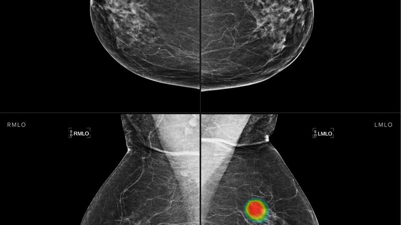
Lunit Insight MMG is once again highlighted in another retrospective study where the associations between the positive predictive values (PPVs) of the abnormality scores generated by an AI-CAD system and the clinical and radiological characteristics of mammograms were evaluated. To focus on suspicious characteristics, only mammograms with abnormal results identified by AI-CAD were included, which resulted in a final dataset with 656 breasts from 599 patients. Results showed that diagnostic indications and positive imaging findings by radiologists were associated with higher abnormality scores. Similarly, the PPVs were significantly higher in women with diagnostic indications, palpability, fatty breasts, and certain imaging findings (masses with or without calcifications and distortion). The work found that the scale of abnormality scores correlated well with the PPVs and that these PPVs values satisfied the BI-RADS recommendations. Additionally, the paper touches upon the need for caution when interpreting AI-CAD scores. Studies like the one presented help radiologists and clinicians better understand the clinical significance of the abnormality scores.
Read full study
Abstract
Objective
Artificial intelligence-based computer-aided diagnosis (AI-CAD) is increasingly used in mammography. While the continuous scores of AI-CAD have been related to malignancy risk, the understanding of how to interpret and apply these scores remains limited. We investigated the positive predictive values (PPVs) of the abnormality scores generated by a deep learning-based commercial AI-CAD system and analyzed them in relation to clinical and radiological findings.
Materials and methods
From March 2020 to May 2022, 656 breasts from 599 women (mean age 52.6 ± 11.5 years, including 0.6% [4/599] high-risk women) who underwent mammography and received positive AI-CAD results (Lunit Insight MMG, abnormality score ≥ 10) were retrospectively included in this study. Univariable and multivariable analyses were performed to evaluate the associations between the AI-CAD abnormality scores and clinical and radiological factors. The breasts were subdivided according to the abnormality scores into groups 1 (10–49), 2 (50–69), 3 (70–89), and 4 (90–100) using the optimal binning method. The PPVs were calculated for all breasts and subgroups.
Results
Diagnostic indications and positive imaging findings by radiologists were associated with higher abnormality scores in the multivariable regression analysis. The overall PPV of AI-CAD was 32.5% (213/656) for all breasts, including 213 breast cancers, 129 breasts with benign biopsy results, and 314 breasts with benign outcomes in the follow-up or diagnostic studies. In the screening mammography subgroup, the PPVs were 18.6% (58/312) overall and 5.1%(12/235), 29.0% (9/31), 57.9% (11/19), and 96.3% (26/27) for score groups 1, 2, 3, and 4, respectively. The PPVs were significantly higher in women with diagnostic indications (45.1% [155/344]), palpability (51.9% [149/287]), fatty breasts (61.2% [60/98]), and certain imaging findings (masses with or without calcifications and distortion).
Conclusion
PPV increased with increasing AI-CAD abnormality scores. The PPVs of AI-CAD satisfied the acceptable PPV range according to Breast Imaging-Reporting and Data System for screening mammography and were higher for diagnostic mammography.